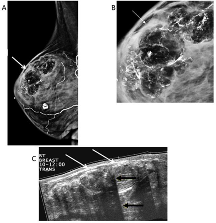
Figure 2.
66-year-old female with left breast cancer, status post-left mastectomy and right reduction mammoplasty. (A) Right MLO view shows multiple circumscribed radiolucent masses with calcifications (white arrows), typical of fat necrosis. (B) Magnified LM view shows the fat contained masses are lined with rim-shell calcifications. (C) Extended field-of-view ultrasound of the upper outer quadrant reveals multiple hyperechoic masses (white arrows) with multiple areas of posterior acoustic shadowing (yellow arrows) related to the curvilinear calcifications. LM, latero-medial; MLO view, mediolateral oblique view.

