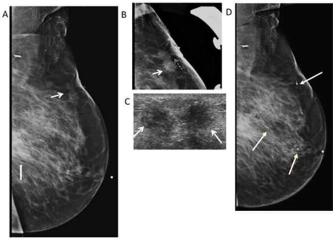
Figure 4.
79-year-old woman with history of breast cancer in 2009 followed by segmental mastectomy and radiation in 2010. She presents for evaluation of palpable abnormality in the left breast. (A) Left MLO mammogram shows a post-surgical scar (thick white arrow) in the central posterior region of the left breast. There is also an irregular non-calcified mass (thin white arrow) with spiculated margins in the superior region of the left breast correlating with the palpable marker (white dotted arrow). (B) Spot LM view shows the spiculated margins of the mass (white arrow) with tethering (white dash arrow) and thickening of the overlying skin. BI-RADS 4C was assigned. (C) Transverse gray- scale ultrasound shows the irregular mass with spiculated margins (white arrows) and heterogeneous hyperechogenicity. Core biopsy was performed revealing fat necrosis. (D) Left MLO view shows multiple clips seen in the left breast related to multiple biopsies of suspicious masses on ultrasound. Pathology shows no malignancy, and recurrent fat necrosis. Bi-RADS, breast imaging-reporting and data system; LM, latero-medial; MLO, mediolateral oblique.

