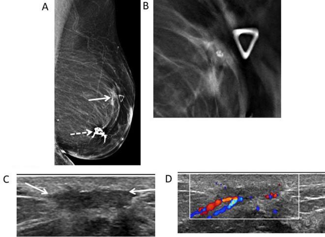
Figure 5.
73-year-old female with history of left breast DCIS, status post-segmentectomy presents for evaluation of palpable abnormality. (A) Left MLO mammogram shows dystrophic calcifications consistent with typical fat necrosis calcifications at the surgical site (white dotted arrow). At the area of palpable abnormality, there is an irregular mass (white arrow). (B.) MLO close-up DBT view allows improved visualization of the spiculated margins of the mass. No entrapped fat was seen. (C) Transverse gray-scale ultrasound shows the superficial hypoechoic mass with irregular margins (white arrows). (D) Color Doppler ultrasound shows the mass demonstrates increased vascularity. BI-RADS: 4C. Core biopsy was performed showing fat necrosis. Bi-RADS, breast imaging-reporting and data system; DBT, digital breast tomosynthesis; DCIS, ductal carcinoma in situ; MLO, mediolateral oblique.

