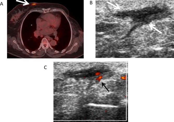
Figure 6.
61-year-old female with left breast cancer followed by mastectomy. (A) Axial fused PET-CT image shows a focal area of increased FDG uptake (white arrow) in the medial region of the right breast with a maximum standardized uptake value of 6.4 suspicious for metastasis or primary breast neoplasm. (B) Transverse gray-scale ultrasound shows an irregular dermal hypoechoic lesion (white arrows) correlating with the lesion shown on the PET-CT. (C) Doppler ultrasound showed internal vascularity (black arrow) within the lesion. BI-RADS 4B. Ultrasound FNA was performed showing fat necrosis. Bi-RADS, breast imaging-reporting and data system; FDG, fluoro-D-glucose; FNA, Fine needle aspiration; PET-CT, positron emission tomography-CT.

