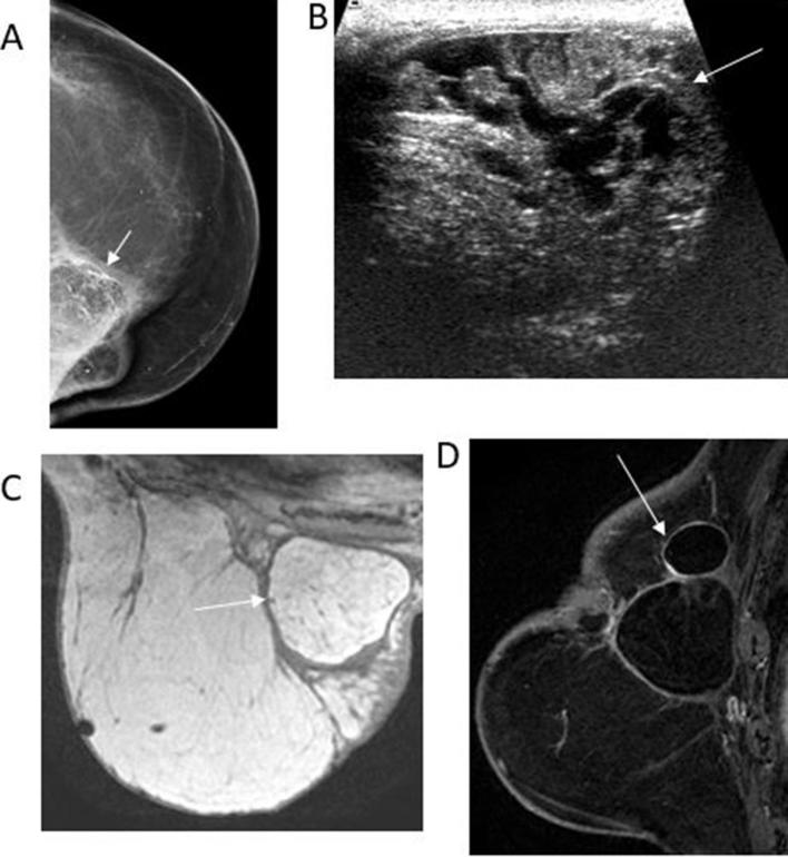
Figure 7.
78-old-year female with history of left breast cancer followed by mastectomy and reconstruction with TRAM flap presented for evaluation of redness and hardness along the medial region of the left breast. (A) Left CC mammographic view reveals lucent masses with peripheral and dystrophic calcifications in the upper inner quadrant (white arrow). (B) Transverse gray-scale ultrasound reveals the mixed echogenicity of the mass (arrow). (C) Axial T1 weighted non-fat-saturated image shows a hyperintense circumscribed mass with a hypointense rim (arrow). The mass signal is similar to the adjacent fat, characteristic of fat necrosis. (D) Sagittal T1 enhanced and fat-suppressed image shows the fat-containing mass with a non-enhancing thin fibrous rim (arrow). Note that no enhancing mass is identified, therefore needle biopsy can be safely avoided. CC, cranial-caudal.

