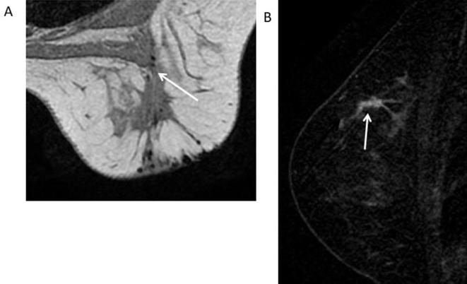
Figure 8.
33-year-old female with history of DCIS followed by breast conservation surgery and radiation. (A) Axial T1 weighted non-fat-suppressed image of the right breast shows a post-surgical scar in the upper outer quadrant (white arrow). (B) Sagittal T1 weighted subtracted image shows a 3 cm clumped enhancement adjacent to the post-surgical scar (white arrow) suspicious for recurrence. BI-RADS 4C. Recommendation: MRI guided biopsy. Core biopsy showed fat necrosis. Bi-RADS, breast imaging-reporting and data system; DCIS, ductal carcinoma in situ.

