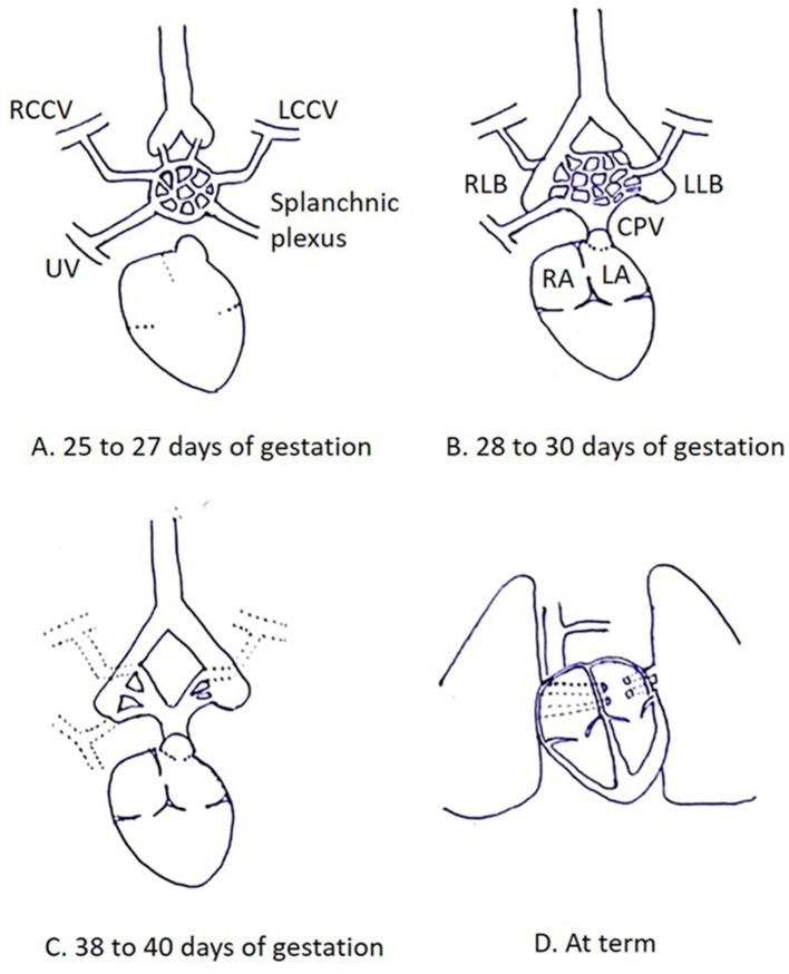
Figure 1.
Normal development of pulmonary venous drainage. (A) Diagram illustrates lung buds draining in to the splanchnic plexus, which in turn communicates with the cardinal and umbilicovitelline venous systems. (B) An outpouching from the dorsal wall of the LA forms the CPV, which communicates with the part of splanchnic plexus draining blood from the primitive lung buds. (C) Connections with cardinal and umbilicovitelline veins involutes. (D) CPV ultimately gets incorporated within the dorsal wall of LA, giving rise to four pulmonary veins which connect directly and separately to the LA. CPV, common pulmonary vein; LA, left atrium; LCCV, left common cardinal vein; LLB, left lung bud; RA, right atrium; RCCV, right common cardinal vein; RLB, right lung bud; UV, umbilical vein.

