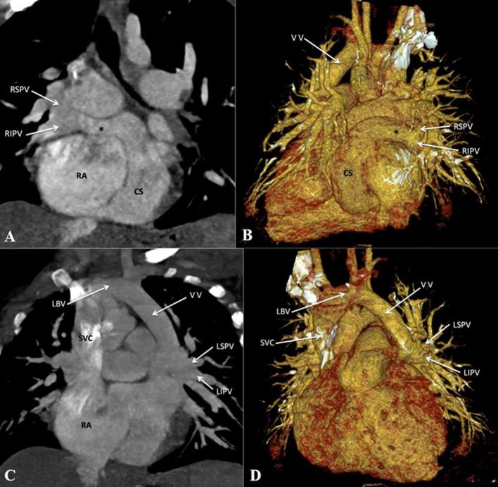
Figure 7.
Mixed total anomalous pulmonary venous connection. (A, B) Contrast-enhanced CT MIP image and volume rendered CT image (posterior view) respectively, show drainage of right pulmonary veins to the dilated CS via a common channel (asterisk). (C, D) Contrast-enhanced CT MIP image and volume rendered image respectively, show the left pulmonary veins draining to the LBV via a VV. CS, coronary sinus; LBV, left brachio cephalic vein; LIPV, left inferior pulmonary vein; LSPV, left superior pulmonary vein; MIP, maximum intensity projection; RIPV, right inferior pulmonary vein; RSPV, right superior pulmonary vein; SVC, superior vena cava; VV, vertical vein.

