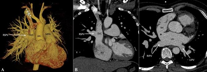
Figure 9.

Partial anomalous pulmonary venous connection. (A) Contrast-enhanced volume rendered CT image (posterior view) and(B) Contrast-enhanced CT MIP image, show drainage of RSPV into the SVC while the other three pulmonary veins drain normally into the LA. C, Axial contrast-enhanced CT MIP image, shows associated sinus venosus atrial septal defect (indicated by asterisk). AA, ascending aorta; LA, left atrium; LIPV, left inferior pulmonary vein; LSPV, left superior pulmonary vein; MPA,main pulmonary artery; RIPV, right inferior pulmonary vein; RSPV, right superior pulmonary vein; superior vena cava.
