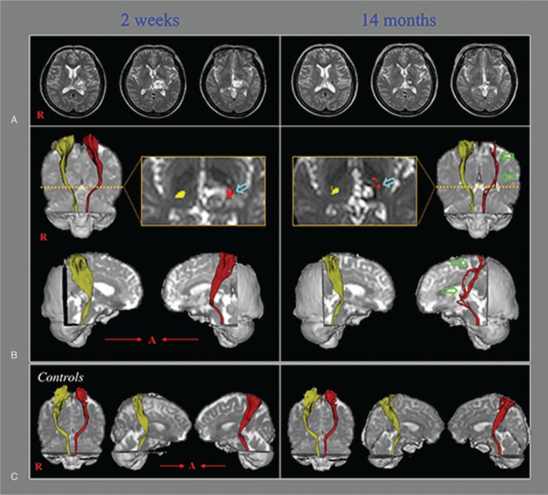
Figure 1.

(A) T2-weighted brain magnetic resonance images showing signs of hemorrhage in the left thalamus (2 weeks after onset) and a leukomalactic lesion in the left thalamus (14 months after onset). (B) Diffusion tensor tractography (DTT) results for the spinothalamic tract (STT). On 2-week postonset DTT, the STT configuration is well-preserved in both hemispheres. However, on 14-month postonset DTT, the left STT reveals partial tearing and thinning (green arrows). The left STT may be seen passing through the area of the thalamic lesion (sky-blue arrows) on both the 2-week and 14-month DTT images. (C) DTT results showing the bilateral STTs in 2 normal subjects (51- and 57-year-old women).
