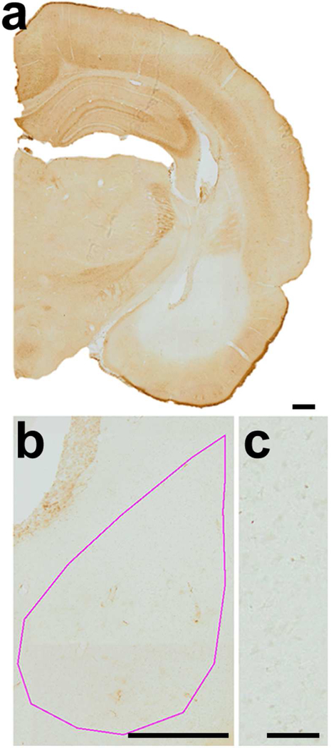
Figure 3. Specificity of WFA labeling of PNN in the BLA of weanling rats.
Panel a: HRP-DAB was used to detect WFA labeling. Specificity of WFA labeling was confirmed by reduction of WFA labeling within the brains injected with the enzymes, chondroitinase-ABC and hyaluronidase, known to dissolve proteoglycans of the PNN and surrounding neuropil. The region infused with chondroitinase-ABC and hyaluronidase exhibited complete absence of the HRP-DAB reaction product, while PNN labeling remained intensely labeled in the surrounding regions (e.g., dorsal hippocampus and reticular thalamus) (scale bar= 200 µm). Panel b: The pink contour indicates the boundary of the BLA (scale bar = 200 µm). Panel c: The lower right panel shows detail of the enzyme-infused region (scale bar = 50 µm). All panels were taken at a magnification of 10X.

