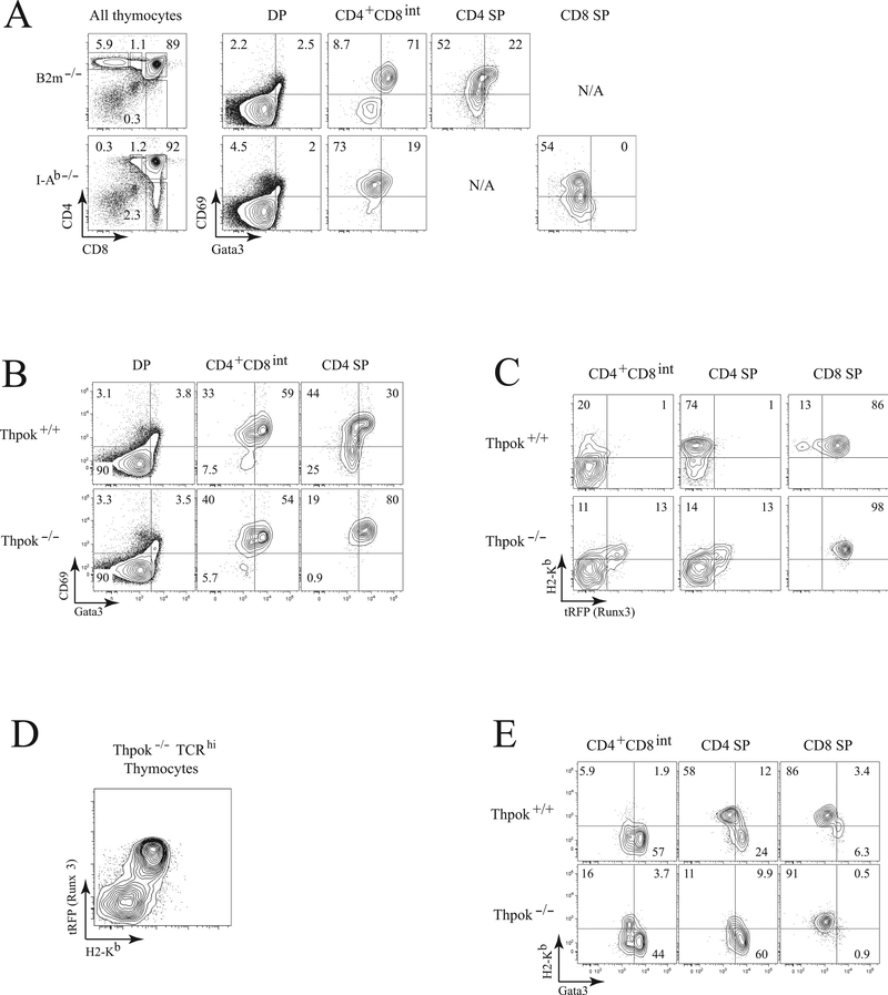
Figure 4. Runx3 and high-level Gata3 expression are mutually exclusive in- thymocytes.
(A) Contour plots (right) show intra-cellular Gata3 and surface CD69 expression in MHC II- (top, from B2m−/− mice) and MHC I-restricted (bottom, from MHC-II-deficient mice) thymocyte subsets as gated on the left. (B) Contour plots show intracellular Gata3 and surface CD69 expression in Thpok+/+ (top) and Thpok−/− (bottom) thymocyte subsets gated as in (A). (C) Expression of H-2Kb and Runx3tRFP in Thpok+/+ and Thpok−/− thymocytes gated as indicated in (A). (D) Plots show surface H-2Kb vs. Runx3tRFP expression in gated TCRhi thymocytes from Thpok−/− mice. (E) Plots show intracellular Gata3 vs. surface H-2Kb expression in CD4+CD8int, CD4 SP and CD8 SP thymocytes from Thpok+/+ and Thpok−/− mice. Data shown are representative of two or more experiments.

