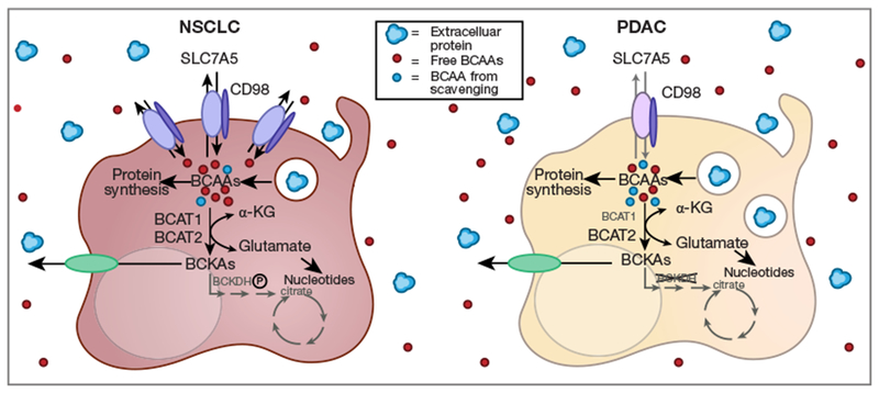
Figure 1.

Differential acquisition of BCAAs in KP NSCLC and KP−/−C PDAC as described by Mayers et al. (16). Relative to normal tissue, SLC7A5 and BCAT1 were over-expressed in NSCLC but not PDAC; BCAT2 protein levels increased in both NSCLC and PDAC. BCKDH was inactivated by phosphorylation in NSCLC while in PDAC BCKDH total protein was greatly reduced. Isotopic labeling studies indicated that carbon from circulating BCAAs ended up in protein and BCKAs in NSCLC to a greater extent than in PDAC. Nitrogen from dietary leucine was used for BCAT-dependent nucleotide synthesis in NSCLC. BSA degradation in the lysosome was increased in isolated PDAC cells relative to NSCLC cells suggesting that PDAC cells may rely on macropinocytosis to compensate for reduced import of extracellular BCAAs through SLC7A5. BCAA, branched chain amino acid; NSCLC, non-small cell lung cancer; PDAC, pancreatic ductal adenocarcinoma; BCKDHA, branched chain keto acid dehydrogenase El alpha; BCAT, branched chain amino acid transferase.
