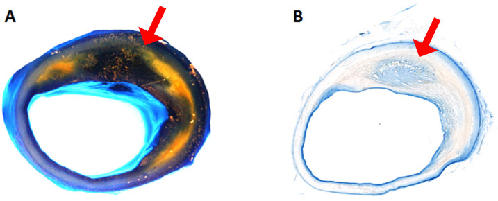
Figure 2. Evans Blue Dye perfused human coronary artery.
A. Gross image of the coronary artery after perfusion. The yellow area is lipid-rich plaque. The dark blue to black are Evans Blue stained areas, see corresponding cryo-sections for blue stain in B (the light blue area is the reflection of the light from luminal surface in A). B. Cryo-sectioned slide from the same artery.

