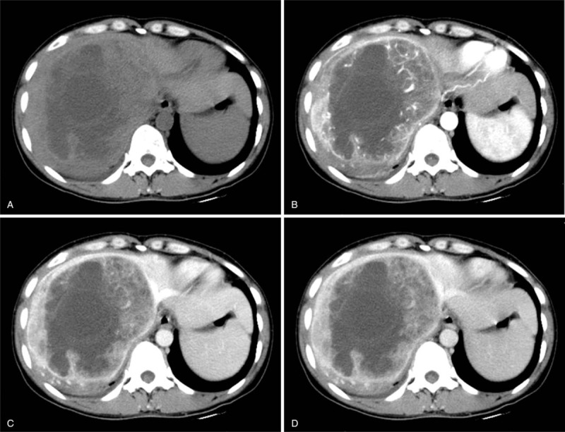
Figure 1.

(A) Nonenhanced computed tomography (CT) scan showing a large heterogeneous tumor of the right liver with ill-defined edges. A contrast-enhanced CT scan indicated significant heterogeneous enhancement of the mass, with multiple wrapped vessels within the wall during the arterial phase (B). The enhancement of solid components decreased during the portal venous phase (C) and delayed phase (D). The unenhanced region of the tumor was confirmed to be necrosis.
