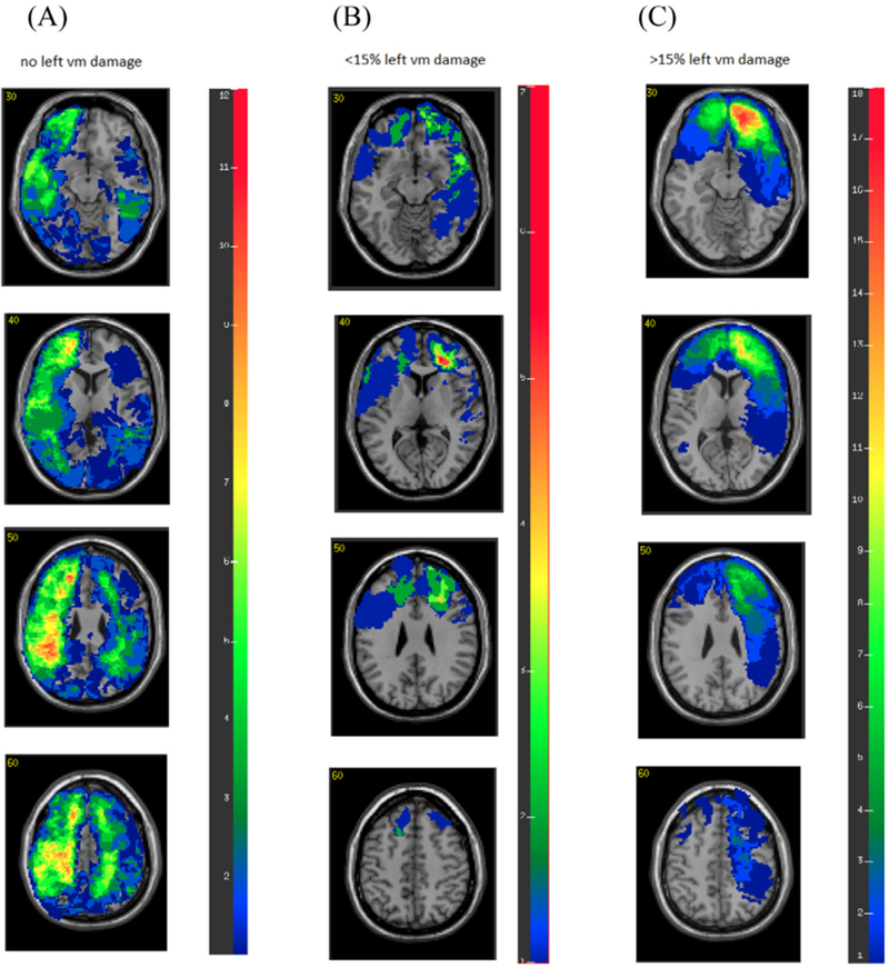
Fig. 2.

Overlay brain lesion maps of pTBI group. (A) Participants with no left vmPFC damage (n = 88). (B) Participants with less than 15% left vmPFC damage (n = 13). (C) Participants with more than 15% left vmPFC damage (n = 18). In each slice, the right hemisphere is on the reader’s left.
