Figure 3.

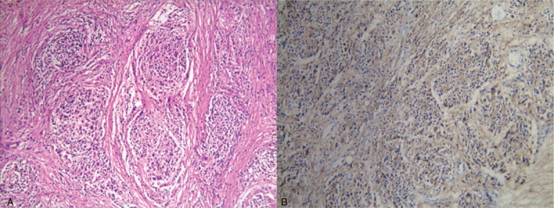
Microscopic images of the surgical specimen. (A) The tumor showed a nested organoid growth pattern (HE, ×100). (B) Immunohistochemical examinations revealed that the tumor cells were positive for Syn (×100). HE = hematoxylin and eosin.

Microscopic images of the surgical specimen. (A) The tumor showed a nested organoid growth pattern (HE, ×100). (B) Immunohistochemical examinations revealed that the tumor cells were positive for Syn (×100). HE = hematoxylin and eosin.