. 2018 Dec 12;7:e41771. doi: 10.7554/eLife.41771

Figure 3. Stability, conformation, and folding kinetics of the template complex.

(A, D, E, I) Extension-time trajectories at constant mean forces with the WT template complex (A) or its variants containing indicated mutations in Munc18-1 (D), VAMP2 (E), or syntaxin (I). The red trace in A shows an exemplary idealized trajectory derived from hidden Markov modeling. Trajectories in A, D, E, and I share the same scale bars. See also Figure 3—figure supplement 1 and Figure 3—source datas 1 and 2 in Dataset 1. (B) Diagram illustrating the transition between the partially closed syntaxin state (state 6 in Figure 2D) and the template complex state (state 7); rates and energies are derived from panel C. (C) Force-dependent unfolding probabilities (top) and transition rates (bottom). Best model fits (solid and dashed curves) reveal the stability and folding and unfolding rates of the template complex at zero force (Figure 4, Table 1, and Figure 3—source data 3 in Dataset 1). (F) Structural model of VAMP2 F77 anchored in the F-pocket in Munc18-1 composed of L247 and T248, which is covered by Y473. The model was derived by superimposing the structures of Munc18-1:syntaxin (Figure 2B; 3C98) and Vps33:Nyv1 (5BV0). (G) Sequence alignment showing F-pocket sequence conservation among SM proteins. (H) Extension-time trajectory of the WT template complex at 5.7 pN. The Qa-R SNAREs were crosslinked between syntaxin L205C and VAMP2 Q36C (Figure 2A, open arrowhead). See also Figure 2—figure supplement 1 and Figure 3—source data 2 in Dataset 1.

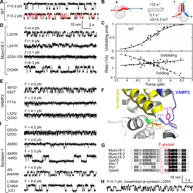
Figure 3—source data 1. MATLAB figure corresponding to Figure 3A with an additional trace at force F = 5.0 pN.
DOI: 10.7554/eLife.41771.019
Figure 3—source data 2. MATLAB figure containing expanded traces shown in Figure 3D,E,I,H.
DOI: 10.7554/eLife.41771.020
Figure 3—source data 3. MATLAB figure for Figure 3C.
DOI: 10.7554/eLife.41771.021

Figure 3.

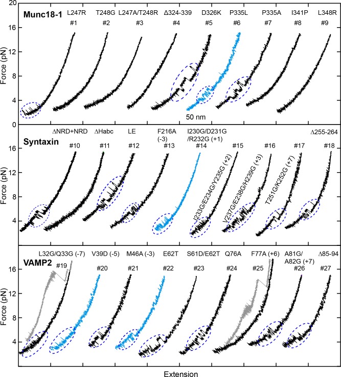
Figure 3—figure supplement 1. FECs obtained in the presence of 2 µM Munc18-1.

Figure 3—figure supplement 1.

Dashed blue ovals mark template complex transitions. Note that the partially closed syntaxin state was abrogated by modifications that are known to destabilize the closed syntaxin, including syntaxin ΔNRD (Burkhardt et al., 2008), the LE mutation (Dulubova et al., 1999; Ma et al., 2011), and Munc18-1 Δ324–339, P335L, and D326K (Munch et al., 2016; Parisotto et al., 2014; Sitarska et al., 2017) (Table 1). For syntaxin LE mutation and I230G/D231G/R232G and Munc18-1 P335A and D326K, template complexes generally formed directly from the open syntaxin (Table 1).
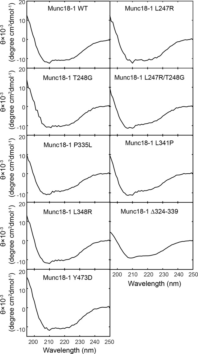
Figure 3—figure supplement 2. Circular Dichroism (CD) spectra show that the mutations tested in our experiments do not significantly alter Munc18-1 folding.

Figure 3—figure supplement 2.

The CD spectra of Munc18-1 mutants that abolished or weakened the template complex are shown, including Munc18-1 F-pocket mutations L247R, T248G, L247A/T248G, disease-related mutations L341P and P335L, phosphomimetic mutation Y473D, and L348R (Parisotto et al., 2014). All the mutant proteins displayed CD spectra closely resembling that of wild-type Munc18-1. In addition, no aggregation at concentrations below 5 mg/ml was noted during the purification of these mutant proteins. These observations suggest that the overall folding of the Munc18-1 proteins are not altered by the mutations. Our results do not however rule out the possibility that some mutations may destabilize the folded protein.
Figure 3—figure supplement 3. Snapshots of the extension-time trajectories at constant mean forces showing sporadic folding of the template complex in the absence of syntaxin NRD.

Figure 3—figure supplement 3.