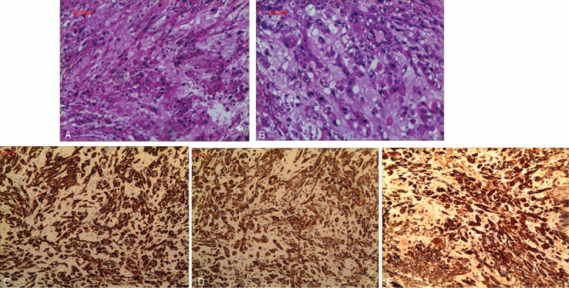
Figure 3.

Histological analysis revealed intracytoplasmic mucin-like substances with large nuclei, deep staining, and an irregular matrix that resembled stroma, cartilage, and mucus with flaky necrosis (A and B). Immunohistochemistry revealed that the specimens were positive for cytokeratin (3+), epithelial membrane antigen (3+), Ki-67 (5%+), S100 (3+), and vimentin (3+) (C, D, and E).
