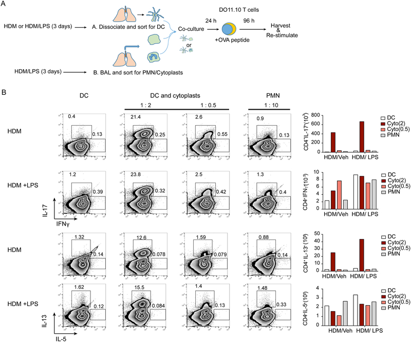
Figure 6: Lung cytoplasts interact with dendritic cells to induce antigen-specific Tlymphocytes.
(a) Schematic diagram showing the antigen-specific T cell activation protocol. Briefly, dendritic cells (DC) were harvested from the lungs of HDM/Veh or HDM/LPS sensitized mice and were incubated overnight with cytoplasts or neutrophils (PMN) at the indicated cell ratios (top of plots) of DC:cytoplasts (1:2 and 1:0.5) and DC:PMN (1:10). The HDM/Veh and HDM/LPS DCs were then co-cultured with naïve CD4+ T cells from DO11.10 mice in the presence of ovalbumin peptide and T cells were re-stimulated and stained for intracellular cytokines (see Methods). (b) Representative flow cytometry plots showing T cells expressing the indicated cytokines. Numbers outside the box represent % of CD4+ T cells. Bar graphs (right) show the absolute cell count for number of cells expressing the indicated cytokine. Data are representative of two experiments.

