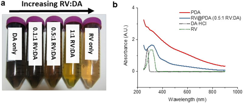
Figure 2:
Optical characterization of PDA and RV@PDA. (a) Appearance of growth solutions containing 0.25 mg/mL DA containing 0.1:1, 0.5:1, and 1:1 RV:DA versus 0.25 mg/mL DA and 0.25 mg/mL RV after 24 h. (b) UV-Vis absorbance spectra of pure PDA and RV@PDA. Spectra of DA and RV only are also shown for comparison.

