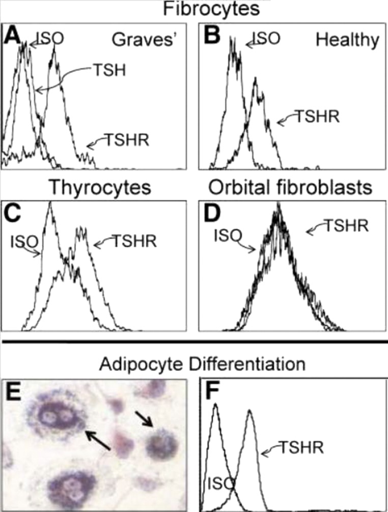
Figure 3.
(A and B) Fibrocytes cultivated from peripheral blood mononuclear cells express high levels of TSHR, regardless of whether they derive from (A) patients with GD or (B) healthy donors. (C) TSHR levels are comparable with those found on primary human thyroid epithelial cells. (D) Undifferentiated OFs fail to express TSHR. (E) Fibrocytes differentiated into adipocytes accumulate intracellular lipid droplets staining with Oil Red O. (F) TSHR levels on fibrocytes remain elevated after differentiation. ISO, isotype control. Reproduced with permission from Douglas RS, Afifiyan NF, Hwang CJ, et al. Increased generation of fibrocytes in thyroid-associated ophthalmopathy. J Clin Endocrinol Metab 2010; 95(1):430-438.

