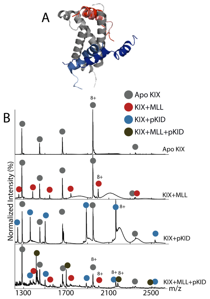
Figure 1.
Structure of KIX with its peptide binding partners and corresponding mass spectra, (A) NMR structures of KIX (gray) with four peptides: MLL (red), E2A (pink), c-Myb (light blue), and pKID (blue). (B) Mass spectra of (from top to bottom) apo KIX, KIX:MLL, KIX:pKID, and KIX:MLL:pKID. The apo peaks are in red, the KIX+MLL peaks in red, KIX+pKID in blue, and the ternary KIX+MLL+pKID in green.

