
Figure 2.
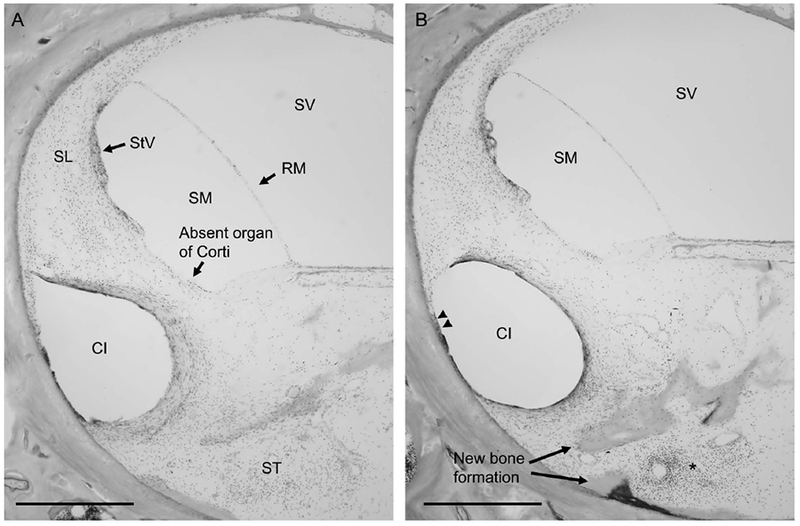
Description of cochlear implant (CI) insertion trauma to spiral ligament in basal turn of the cochlea.Figure 2A, The impression of the CI electrode migrates at the anterior curvature of the cochlea to occupy the spiral ligament, which is confluent with the loose areolar tissue of the scala tympani (ST). Note the absent organ of Corti in this region of the basal turn of the cochlea. Figure 2B, CI is seen to erode through the endosteal layer of the bone at the superior anterior portion of the basal turn (double arrowheads), with evidence of suppuration (asterisk) surrounded by new bone formation (arrowheads).
SL = spiral ligament, RM = Reissner’s membrane, StV = stria vascularis, SM = scala media, ST = scala tympani, SV = scala vestibuli, CI = cochlear implant electrode impression, double arrowheads = erosion of CI electrode through endosteal layer of the bone into marrow spaces, arrows = new bone formation. Scale bar, 500 μm.