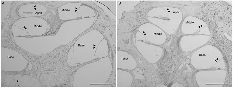
Figure 3.

Presence of endolymphatic hydrops in the patient with a multichannel cochlear implant (3A, LEFT) with comparison to a normal hearing patient (3B, RIGHT). Mid-modiolar cross-sections, low-magnification light microscopy images, magnification 2.5x; Scale bar, 1mm. Double arrowheads point the location of Reissner’s membrane. Figure 3A, Right ear of our patient with a multichannel cochlear implant. Note the presence of endolymphatic hydrops (bulging of Reissner’s membrane with expansion of the scala media with the double arrowheads pointing to Reissner’s membrane) in the apical and middle turns with less noticeable but present endolymphatic hydrops in the basal turn. Area of fibrosis around the cochlear implant electrode denoted by the asterisk. Figure 3B, Comparison image from a patient with normal hearing and normal caliber of the scala media.
