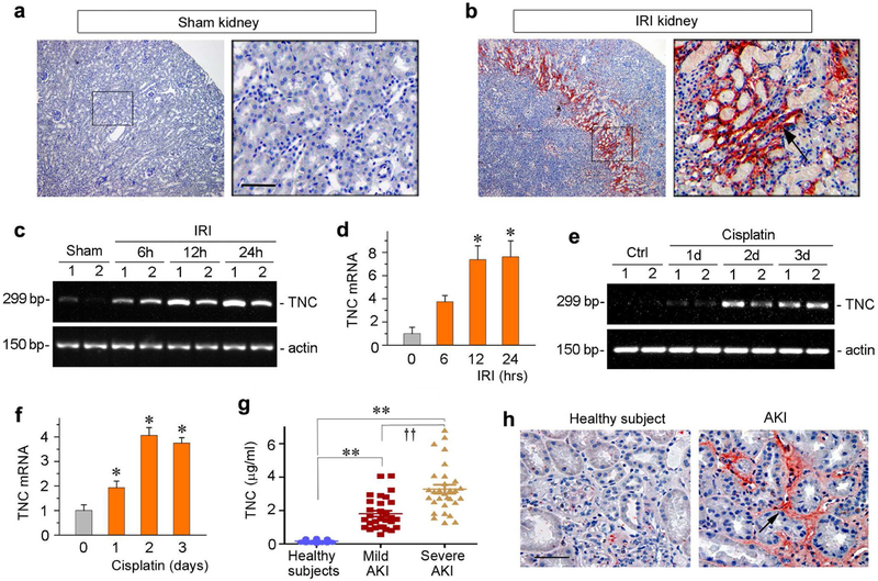
Figure 1. Expression of TNC is induced in animal models of AKI and in humans.
(a, b) Representative micrographs show TNC protein expression in sham and ischemic kidney at 30 hours after IRI. Kidney sections were stained immunohistochemically with specific antibody against TNC. Boxed area was enlarged. Arrow indicates positive staining. Scale bar, 50 μm. (c, d) RT-PCR results show the relative mRNA abundances of TNC at different time points after IRI. Representative RT-PCR results (c) and quantitative data (fold induction over the controls) (d) are presented. *P < 0.05 versus controls (n = 5 to 6). (e, f) RTPCR results show the relative mRNA abundances of TNC at different time points after cisplatin. Representative RT-PCR results (e) and quantitative data (fold induction over the controls) (f) are presented. *P < 0.05 versus controls (n = 5 to 6). (g) The circulating levels of TNC are elevated in human AKI. TNC protein was detected by a specific ELISA in the plasma of normal healthy subjects and patients with mildand severe AKI after cardiac surgery, respectively. **P< 0.01 versus healthy subjects; ††P< 0.01 versusmild AKI. (h) Representative micrographs show the abundance and localization of TNC proteins in healthy control and AKI patients as indicated. Scale bar, 50 μm.

