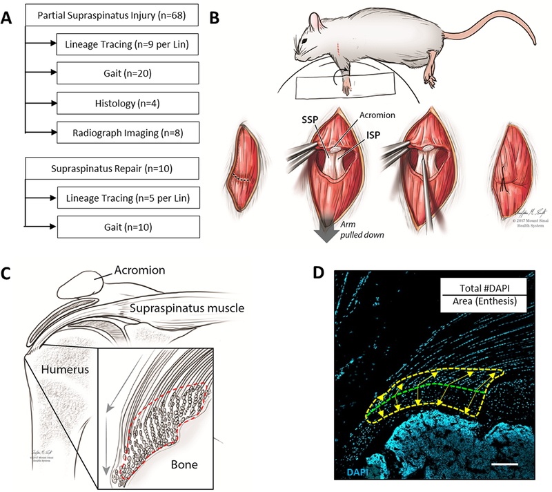
Figure 1: Experimental design and methods overview.
(A) Experimental design and sample size for supraspinatus injury models. (B) Schematic demonstrating partial supraspinatus tendon tear surgery. (C) Schematic and (D) DAPI-stained histological section of the supraspinatus tendon defining the enthesis region of interest used for cell quantification (dotted lines). Transition between mineralized and unmineralized fibrocartilage is indicated by the green dotted line and equidistant points extending toward bone and tendon used to define the outer boundaries of the region of interest (yellow arrows). Scalebar: 100μm.

