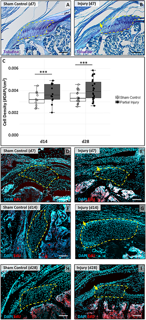
Figure 3: Loss of enthesis architecture and hypercellular scar formation after partial supraspinatus tear.
(A, B) Toluidine blue staining of supraspinatus tendon sections show loss of enthesis structure in the localized defect by d7. (C) Quantification of DAPI stained sections show increase in cell density at d14 and d28 (n=12–22). *** indicates significant difference (p<0.001). DAPI and EdU detection of supraspinatus tendon sections show very limited cell proliferation in the scar at (D, E) d7 and no proliferation at (F, G) d14 or (H, I) d28. Localized enthesis scar indicated by yellow arrows. Region of interest highlighted by yellow dashed outlines. Scalebars: 100um.

