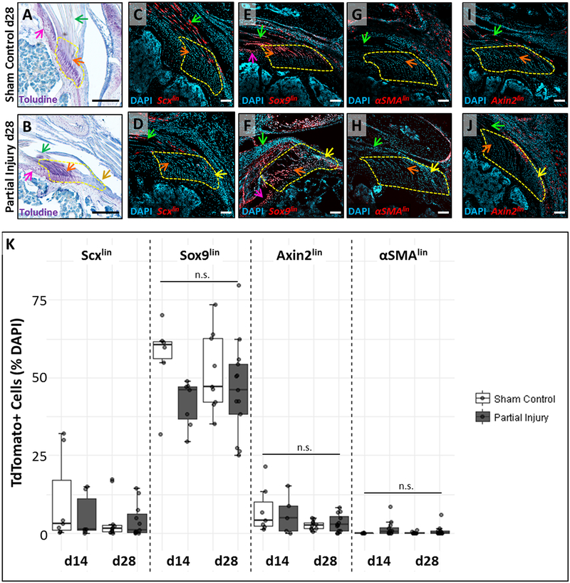
Figure 4: Lineage tracing does not identify the source of enthesis scar cells.
(A, B) Toluidine blue staining of supraspinatus tendon sections show sustained loss of enthesis structure in the localized defect at d28. Lineage tracing using (C, D) ScxCreERT2, (E, F) Sox9CreERT2, (G, H) αSMACreERT2, and (I, J) Axin2CreERT2 did not show observable contribution of TdTomato+ cells to the scar defect. (K) Quantification of %TdTomato cells in the region of interest. n.s. indicates not significantly different (p>0.05). Arrows indicate: tendon (green), un-mineralized enthesis fibrocartilage (orange), scar (yellow), and articular cartilage (pink). White scalebars: 100um. Black scalebars: 200um.

