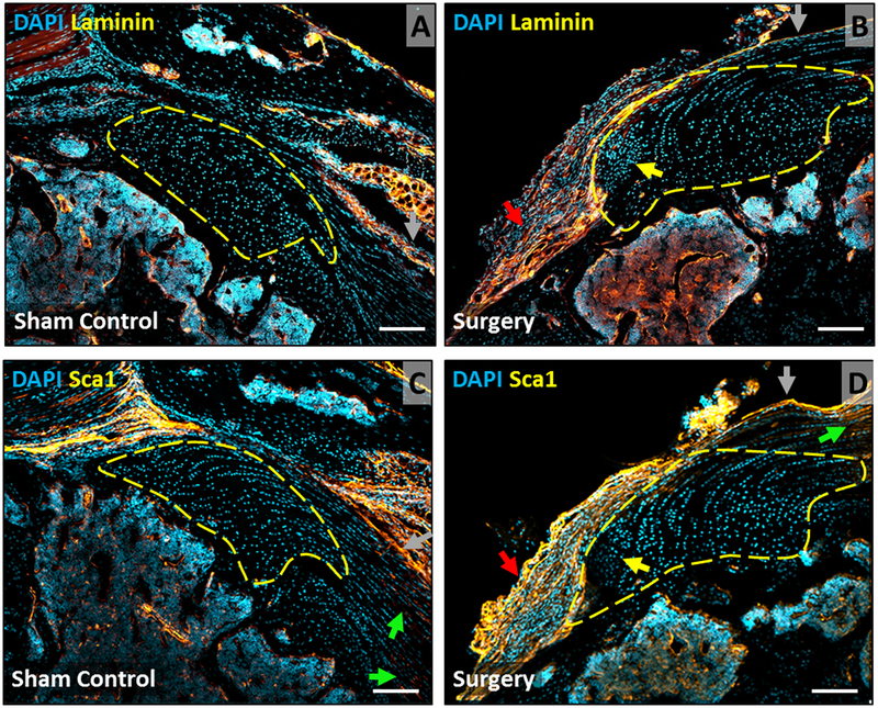
Figure 5: Laminin and Sca1 immunostaining shows expansion of bursa at d28 after injury.
Immunostaining for (A, B) laminin and (C, D) Sca1 in supraspinatus tendon sections at d28 highlight epitenon, bursa, and bone marrow cells, with no staining of enthesis fibrocartilage or scar cells. Arrows indicate: enthesis scar (yellow), bursa (red), tendon (green), epitenon (gray). Scalebars: 100um.

