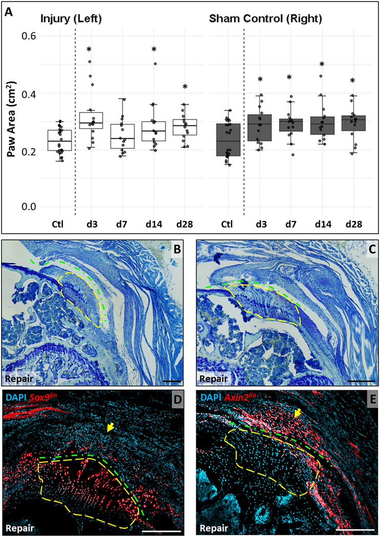
Figure 6: Full tendon detachment and repair results in functional changes and recruitment of Axinlin scar cells.
(A) Paw area is generally significantly increased in both limbs of injured animals compared to limbs of non-injured animals. (B, C) Toluidine blue staining of supraspinatus tendon sections show disruption of enthesis organization and characteristic orientation with massive scar formation. Lineage tracing using (D) Sox9CreERT2 shows TdTomato+ cells in enthesis fibrocartilage with no labeling in the scar. (E) Axin2CreERT2 tracing shows little labeling of enthesis fibrocartilage but considerable presence of TdTomato+ cells in the scar. Yellow dashed outlines enclose similar regions. Green dashed line indicates detachment site. Black scalebars: 200μm. White scalebars: 100μm.

