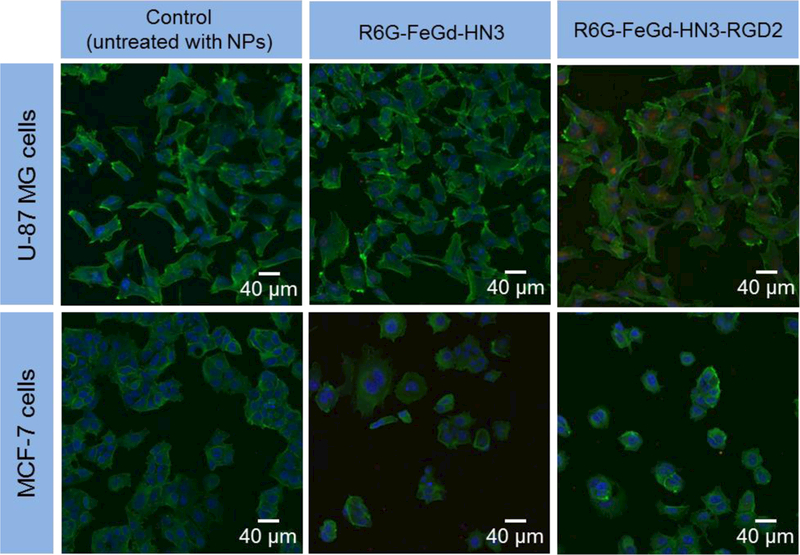
Figure 4.

LSCM images of U-87 MG or MCF-7 cells incubated with R6G-FeGd-HN3 or R6G-FeGd-HN3-RGD2. The cells untreated with nanoparticles are used as the control. The cytoskeleton stained with phalloidin-FITC is green and the nucleus stained with Hoechst is blue. The R6G-loaded nanoparticles are red.
