Fig. 3.
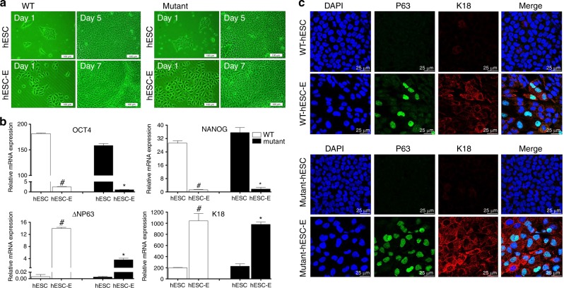
Direct differentiation of human embryonic stem cells (hESCs) into epithelial progenitor cells (hESC-Es). a Morphological changes in wild-type (WT) (PTCH1+/+) and mutant (PTCH1R135X/+) hESCs before and after epithelial induction, as assessed by inverted phase contrast microscopy. Scale bar = 100 μm. b Quantitative real-time PCR analysis of the expression of pluripotent markers (OCT4, NANOG) and keratinized epithelial markers (K18, ΔNP63) in undifferentiated hESCs and induced hESC-Es. Data represent the mean ± SD, n = 3 (#, WT hESC-Es vs WT hESCs; *, mutant hESC-Es vs mutant hESCs; #, *, P < 0.05). c Immunofluorescence images of P63 (green) and K18 (red) expression in WT and mutant cells before and after induction. The left panel displays corresponding DAPI nuclear staining (blue) and the right panel displays corresponding merged images. Scale bar = 25 μm

