Fig. 1.
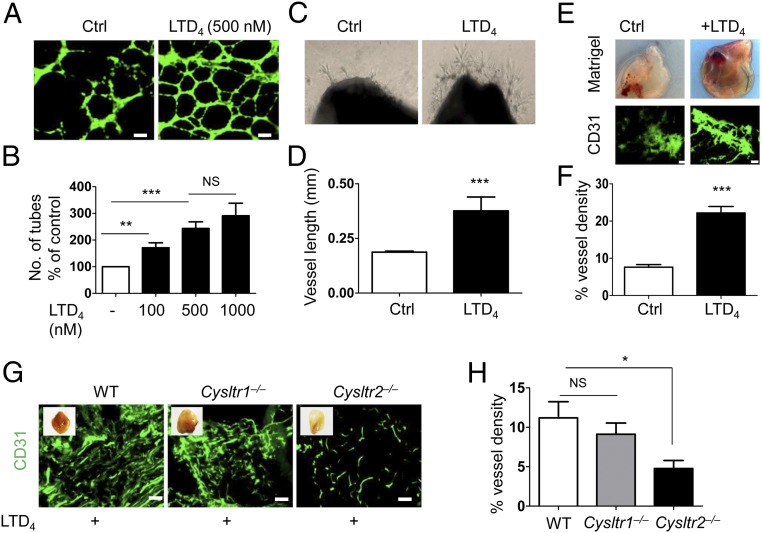
LTD4 induces angiogenesis through CysLT2R. (A and B) MDECs were plated on growth factor-reduced Matrigel and treated with or without LTD4 (100, 500, and 1,000 nM) for 12–16 h. Cells were stained with calcein AM and fixed. (A) Representative images of tube formation with and without LTD4 (500 nM) treatment. Ctrl, control. (Scale bars: 200 μm.) (B) Quantification of tube formation. Tubes from at least 10 representative images of each replicate were counted manually and presented as the average percentage from three separate experiments. (C) Representative photographs of aortic rings from WT mice placed in growth factor-reduced Matrigel and then treated with or without LTD4 for 7 d. (D) Quantification of sprout length (n = 6–8). (E–H) Growth factor-reduced Matrigel supplemented with bFGF (200 ng/mL) alone (Ctrl) or in the presence of LTD4 (500 nM) was injected into both flanks of indicated mouse groups for 21 d. (E, Upper) Representative photographs of Matrigel plugs. (E, Lower) Matrigel sections immunostained for CD31. (Scale bars: 100 μm.) (F) Quantification of CD31 fluorescence (n = 10 per group) from E. (G) Representative CD31-stained Matrigel plug sections from the indicated mouse groups. (Scale bars: 100 μm.) (H) Quantification of CD31 staining from G (n = 8–12 per group). Data are shown as mean ± SEM. *P < 0.05; **P < 0.01; ***P < 0.001; NS (nonsignificant) as determined by the Mann–Whitney U test (B, D, and F) and one-way ANOVA with a Tukey post hoc test (H).

