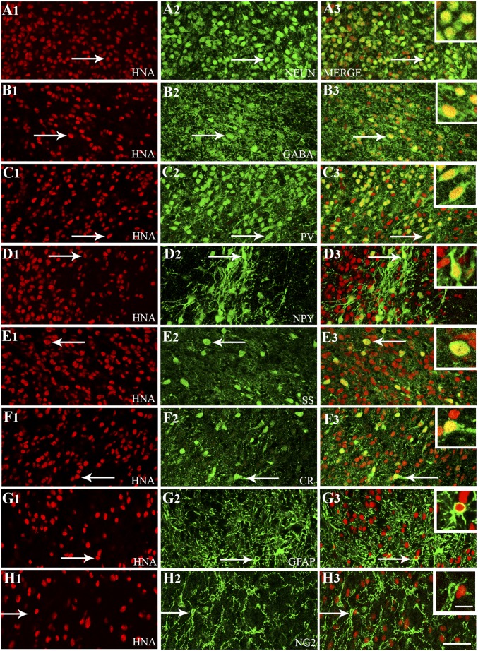
Fig. 5.
Cells derived from hMGE-like cell grafts placed into the hippocampus after SE differentiated predominantly into GABA-expressing interneurons comprising various subclasses. Differentiation of hMGE graft-derived cells into neurons expressing neuron-specific nuclear antigen (NeuN; A1–A3) and interneurons expressing GABA (B1–B3) is illustrated. Differentiation of hMGE graft-derived cells into subclasses of interneurons expressing PV (C1–C3), NPY (D1–D3), SS (E1–E3), and calretinin (CR; F1–F3) is illustrated. (G1–H3) Differentiation of hMGE graft-derived cells into GFAP+ astrocytes is minimal, and none of the hMGE graft-derived cells differentiate into neuron-glia 2+ (NG2+) oligodendrocyte progenitors. All cells in red (first column) denote graft-derived cells expressing HNA (a marker of human cells), whereas cells in green (second column) illustrate cells expressing neuronal or glial antigens. The third column illustrates merged images from columns 1 and 2. Arrows in A1–G3 denote examples of dual-labeled cells, whereas arrows in H1–H3 denote a host NG2+ cell. (Insets) In the third column, magnified views of cells indicated by arrows are displayed. (Scale bars: A1–H3, 50 μm; Insets, 20 μm.)

