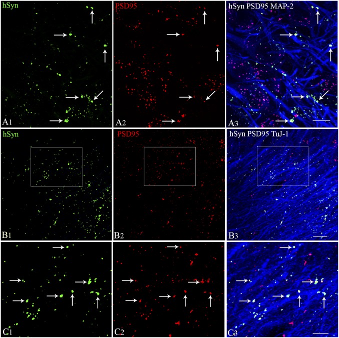
Fig. 8.
Synapse formation between graft-derived axons and host hippocampal excitatory neurons in the DG and the CA1 subfield. Synapses between hSyn-expressing axon endings from graft-derived neurons (A1, green) and PSD-95–expressing regions (A2, red) on MAP-2 positive dendrites of DG granule cells (A3, blue) are illustrated. Direct contacts between hSyn+ and PSD-95+ structures show the location of synapses on DG granule cell dendrites (indicated by arrows in A3). Synapses between hSyn+ axon endings from graft-derived neurons (B1, green) and PSD-95+ regions (B2, red) on TuJ-1+ dendrites of CA1 pyramidal neurons (B3, blue) are illustrated. (C1–C3) Magnified views of areas from B1–B3 showing synaptic contacts between graft-derived hSyn+ presynaptic terminals on PSD95+ postsynaptic regions in the host hippocampal CA1 pyramidal neuron dendrites. Two 1-μm-thick consecutive optical sections (two adjacent sections) were employed to confirm the presence of both pre- and postsynaptic puncta (hSyn- and PSD95-stained structures) on dendrites. (Scale bars: A1–A3 and C1–C3, 10 μm; B1–B3, 20 μm.)

