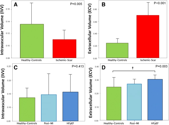
Fig. 2.
Top Panel: Bars and 95% confidence intervals of intravascular (IVV; a) and extracellular (ECV; b) volumes in healthy controls (green) and ischemic scar (red). IVV was reduced and ECV was strongly augmented in the ischemic scar as compared to normal myocardium. Bottom Panel: Bars and 95% confidence intervals of IVV (c) and ECV (d) in healthy (green), post-MI patients (remodeled myocardium not including the scar, light blue) and HFpEF patients (blue). Bonferroni’s post-hoc analysis, † P < 0.05

