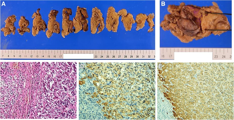
Fig. 3.
A left adrenal gland tumor removed by adrenalectomy. a, b Gross pathology of the tumor of the left adrenal gland. c–e Microscopic image of the tumor under hhematoxylin and eosin staining (c), chromogranin A immunostaining (d), or synaptophysin immunostaining (e). The magnification was 200×. d, e The cells stained in brown are positive for chromogranin A and synaptophysin, respectively

