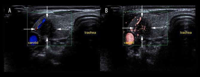
Figure 4.
Evaluation of vascularity in a lesion located in the right thyroid lobe on color Doppler flow imaging (CDFI) and superb microvascular imaging (SMI) in a 31-year-old woman. (A) Color Doppler flow imaging (CDFI) shows peripheral vascularization on the top left of the thyroid nodule (grade 1). (B) Superb microvascular imaging (SMI) shows both peripheral and central vascularity (grade 2) with some linear and branching small vessels. The thyroid nodule was diagnosed histologically as a papillary thyroid carcinoma.

