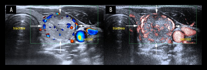
Figure 5.
Color Doppler flow imaging (CDFI) and superb microvascular Imaging (SMI) show increased vascularity in a lesion located in the left thyroid lobe in a 25-year-old woman. (A) Color Doppler flow imaging (CDFI) shows peripheral and central vascularization (Grade 2) with linear and dot-like vessels. (B) Superb microvascular imaging (SMI) shows peripheral and central vascularity (Grade 3) with more linear and branching small vessels. The thyroid nodule was diagnosed histologically as a follicular adenoma.

