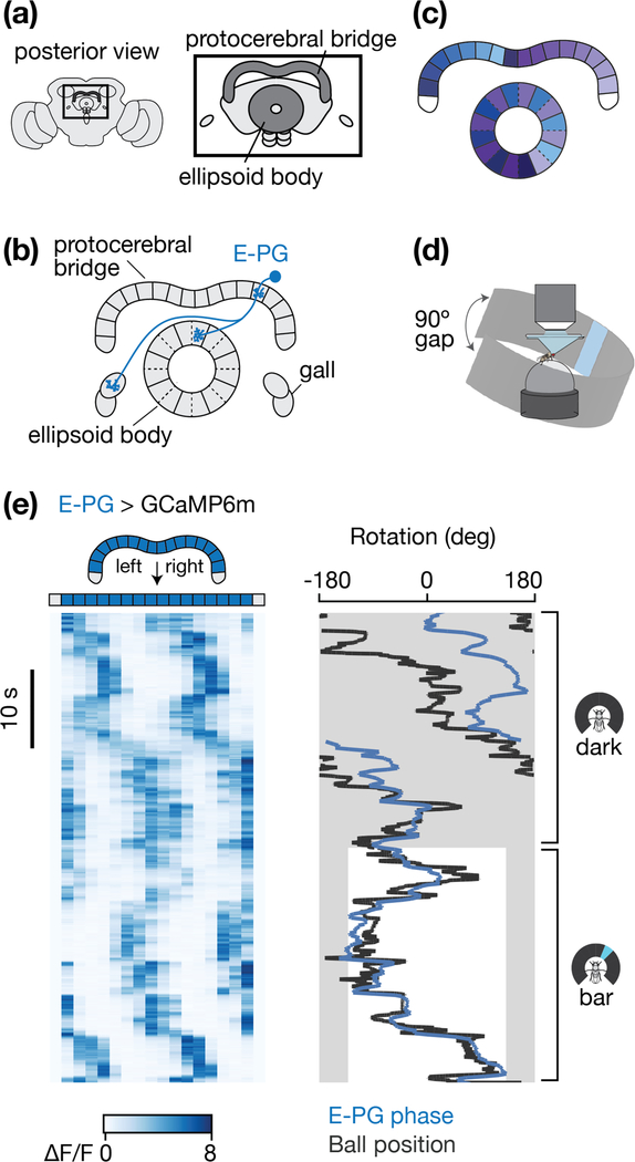
Figure 1 |. A heading signal in the fly central complex.
a, The protocerebral bridge and ellipsoid body represent two structures in the central complex, a set of interconnected neuropils located in the middle of the fly brain. b, The anatomy of one E-PG neuron is shown. Glomeruli in the protocerebral bridge and tiles in the ellipsoid body are delineated by solid lines. Wedges in the ellipsoid body are delineated by dashed lines. c, The full array of E-PGs innervate the entire ellipsoid body and most of the bridge. For E-PGs, each half of the bridge maps to every second wedge in the ellipsoid body. Colors represent the mapping of E-PG neurons between the ellipsoid body and bridge. d, Preparation for imaging a fly walking on an air-supported spherical treadmill with a panoramic LED display. The LED display covered 270˚ around the fly, with a 90˚ gap in the back (arrows) e, (Left) E-PG activity in the protocerebral bridge as the fly walks in complete darkness (top) and as it walks with a tall, bright, vertical bar presented on the LED display, which rotates in closed-loop with the fly’s turning to indicate its virtual heading. (Right) The position, or phase, of the E-PG activity in the bridge (blue) and the ball’s yaw orientation (black). We shifted the E-PG phase (blue curve) by a constant offset so as to best match the ball/bar position (black curve) when the bar is visible. Grey areas highlight the 90º gap in the arena behind the fly where the bar is not visible.

