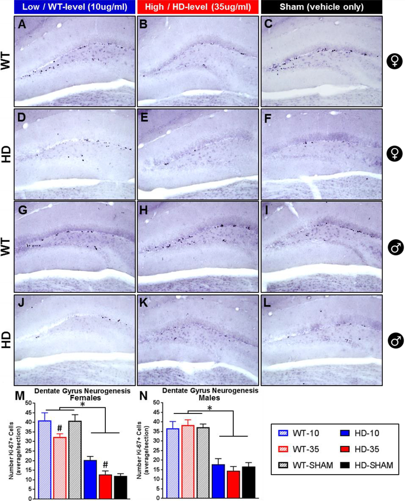
Figure 6. Effect of glucocorticoid replacement on neurogenesis in the dentate gyrus.
Representative images of Ki-67+ mitotic cells, a marker for neurogenesis, in the dentate gyrus of wild-type (females A-C, males G-I) and transgenic R6/2 HD mice (females D-F, males J-L). R6/2 mice show fewer ki67+ cells than WT (M,N - p<.05) and treatment with low dose CORT (WT-level, 10μg/ml) increases ki67+ cells in females relative to those on high dose (HD-level, 35μg/ml) (M, Treatment: p<.05, # Tukey’s post-hoc indicates difference between 10μg/ml and 35μg/ml treated female mice, regardless of genotype). Treatment had no effect on males (N, p>.05). N=24 for each sex, n=4 per group. Values represent Mean ± SE.

