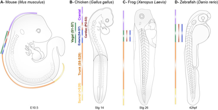
Figure 1. The anterior-to-posterior regionalization of the neural crest.
Neural crest cells are regionalized into four major anterior-posterior axial levels along the dorsal neural tube of vertebrate embryos. Approximate boundaries of cranial (purple), vagal (green), trunk (orange), and sacral (yellow) neural crest populations are indicated for mouse (E10.5), chick (Hamburger-Hamilton 14), frog (Nieuwkoop-Faber 26), and zebrafish (42hpf) embryos. The cardiac (red) and enteric (blue) subpopulations of the vagal neural crest are also indicated across species.

