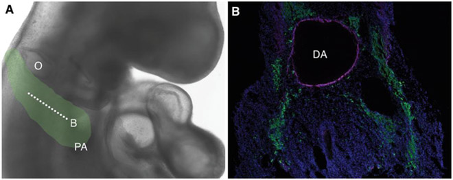
Figure 2. Cardiac neural crest migration.
(A) Cardiac neural crest cells derived from the post-otic region migrate towards the pharyngeal arches in a stage HH18 chick embryo. Green shading in (A) indicates path of cardiac neural crest. Cross section in (B) is indicated by dotted line. (B) Transverse section through a stage HH18 embryo shows neural crest immunostained for HNK-1 (green) migrating around the dorsal aorta (DA) to enter the pharyngeal arches. Nuclei are labeled with DAPI (blue); immunostaining for smooth muscle actin (magenta) lines the DA. O, otic; PA, pharyngeal arches; DA, dorsal aorta.

