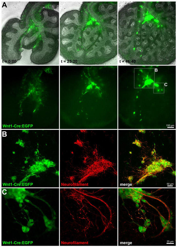
Figure 3. Lineage tracing reveals vagal neural crest migration into mouse lung buds.
(A) E11.5 Wnt-1Cre/ZEG mouse lungs were explanted and imaged at 20-minute intervals for 46 hours and 40 minutes, displaying extensive neural crest cell migration and coalescing into pulmonary ganglia. See also Supplemental Movie 1. (B-C) Explanted lungs were processed for immunostaining against neurofilament M, and imaged by confocal microscopy. EGFP positive cells extend a complex network of neurofilament positive fibers interconnecting the coalescing ganglia. (Images acquired at the 2014 Marine Biological Laboratory Embryology course, with the assistance of Dr. Angelo Iulianella and the mouse module).

