Figure 5.
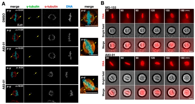
Rotation of mitotic spindle in A83-01-treated cells. (A) After incubation with RO-3306 for 20 h, cells were released in the presence of 3 µM A83-01 for 1 h, fixed, and stained for α-tubulin, γ-tubulin and DNA. (Left), z–stack images were acquired by using a confocal microscopy, and one or two focal planes (x-y images) were shown. (Right), the x–z projections from the z–stack images of 25 focal planes (1 µm apart). Arrows indicate the positions of centrosomes. Scale bars, 10 µm. (B) After incubation with RO-3306, cells were released in the presence of 0.1 µM Hoechst 33342 with 10 µM MG-132 or 3 µM A83-01. MG-132 or A83-01 was added into the culture at the time of release or at 30 min after the release, respectively. Then, mitotic progression was monitored every 5 min by time-lapse imaging until 3 h after the release. Fluorescence of Hoechst 33342 and bright field images are shown. Scale bars, 10 µm.

