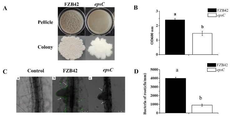
Figure 6.
Biofilm formation by wild type FZB42 and the epsC mutant. The upper panel illustrates the formation of a pellicle by cells cultured at 30 °C for three days on LBGM medium (Lysogeny broth supplemented with 1% (v/v) glycerol and 0.1 mM MnSO4); the lower panel illustrates colony morphology (A), quantitative spectrophotometric assay of biofilms stained with crystal violet (B), confocal laser scanning microscopy analysis of root colonization. Scale bar, 100 µm (C), adherence capacities of wild type and epsC mutant to the A. thaliana root (D). Values in (B) and (D) are shown as means, with the whiskers representing the standard error (SE, n = 12). Different letters above each column indicate statistically significant (p < 0.05) differences in mean performance.

