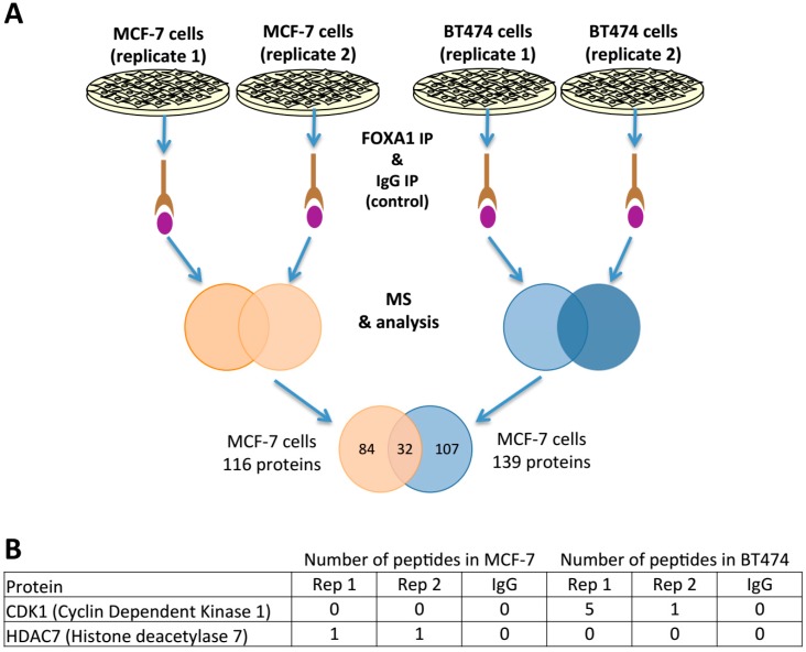
Figure 4.
Proteomics results from FOXA1 immunoprecipitation (IP). (A) Workflow for FOXA1 IP proteomics. Two independent replicates were performed and uniquely proteins without peptides identified at IgG control were considered positive. Moreover, we considered exclusively proteins identified in both of the replicates. The figure includes a Venn diagram that compares the number of FOXA1 interacting proteins shared between MCF-7 and BT474 cells and the ones identified exclusively each cell line tested. (B) Peptide enrichment of CDK1 and HDAC7 in both cell lines is included. The rest of peptide enrichment of the other identified proteins can be found at supplementary information.

