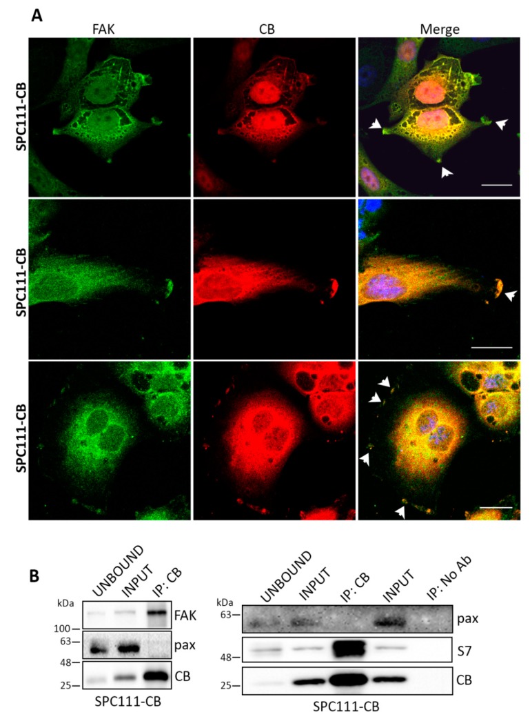
Figure 3.
CB co-localizes with focal adhesion kinase (FAK) at focal adhesion sites. Co-IP experiments confirm an interaction of CB with FAK and septin 7. (A) Representative confocal images from fixed cells stained for FAK (green) and CB (red) in SPC111-CB cells. CB co-localizes with FAK at the leading edge of the cells and forms dot-like patterns (upper and bottom panels) and membrane ruffles (middle and bottom panels) typical of focal adhesions (see arrowheads) as previously shown for CR [13]. Scale bar: 20 µm; (B) Co-IP experiments with cellular lysates from SPC111-CB cells shows co-immunoprecipitation of CB with FAK and septin 7. An input sample collected before immunoprecipitation is shown as INPUT. The INPUT, UNBOUND, as well as the immunoprecipitated (IP) samples were separated using 10% SDS-PAGE and followed by Western blot analysis using anti-CB, anti-FAK, anti-septin 7 and anti-paxillin (negative control) antibodies (n = 3 independent experiments). A co-IP with no antibodies is shown as a negative control of the assay.

