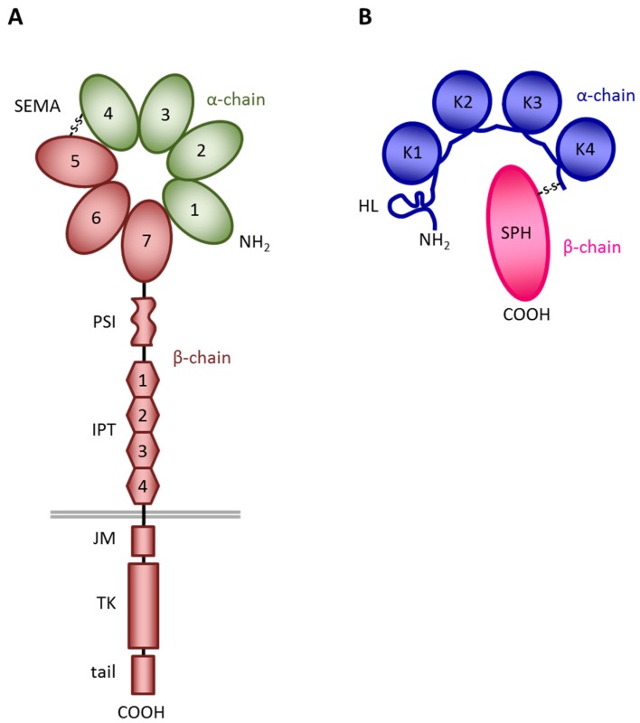
Figure 1.
Schematic structure of MET and HGF. (A) MET structure. SEMA, a seven-bladed β-propeller domain shared with Semaphorins and Plexins; PSI, a cysteine-rich domain present also in Plexins, Semaphorins, and Integrins; IPT, four immunoglobulin-like domains present also in Plexins and Transcription factors; JM, juxtamembrane domain, a region containing residues that act as negative regulators of the kinase activity; TK, tyrosine kinase domain endowed with enzymatic activity; tail, a C-terminal domain containing the multifunctional docking site. In green the α-chain, in brown the β-chain of MET. The grey line represents the cell membrane. (B) HGF structure. HL, hairpin loop; K1–K4, kringle domains; SPH, serine-protease homology domain, devoid of protease activity. In blue the α-chain, in red the β-chain of HGF. S-S, disulphide bond linking the α- and β-chains of the two molecules.

